Saturday, 12 November 2016

TOTAL HIP REPLACEMENT

TOTAL HIP REPLACEMENT - A total hip replacement is a surgical procedure whereby the diseased cartilage and bone of the hip joint is surgically replaced with artificial materials. The normal hip joint is a ball and socket joint. The socket is a "cup-shaped" component of the pelvis called the acetabulum. The ball is the head of the thighbone. Total hip joint replacement involves surgical removal of the diseased ball and socket and replacing them with a metal ball and stem inserted into the femur bone and an artificial plastic cup socket. The metallic artificial ball and stem are referred to as the "femoral prosthesis" and the plastic cup socket is the "acetabular prosthesis." Upon inserting the prosthesis into the central core of the femur, it is fixed with a bony cement called methylmethacrylate. Alternatively, a "cementless" prosthesis is used that has microscopic pores which allow bony ingrowth from the normal femur into the prosthesis stem. This "cementless" hip is felt to have a longer duration and is considered especially for younger patients. Total hip replacement is also referred to as total hip arthroplasty.


Total hip replacements are performed most commonly because of progressively worsening of severe arthritis in the hip joint. The most common type of arthritis leading to total hip replacement is degenerative arthritis of the hip joint. This type of arthritis is generally seen with aging, congenital abnormality of the hip joint. The progressively intense chronic pain, together with impairment of daily function including walking climbing stairs, and even arising from a sitting position, eventually become reasons to consider a total hip replacement. Because replaced hip joints can fail with time, whether and when to perform total hip replacement are not easy decisions, especially in younger patients.

Monday, 7 November 2016

TOTAL KNEE REPLACEMENT

TOTAL KNEE REPLACEMENT - Patients with severe destruction of the knee joint associated with progressive pain and impaired function maybe candidates for total knee replacement. Osteoarthritis is the most common reason for knee replacement operation. Physical therapy is an essential part of rehabilitation after total knee replacement. A TKR is a complex procedure that requires an orthopedic surgeon to make precise measurements and skillfully remove the diseased portions of your bone, in order to shape the remaining bone to accommodate the knee implant. After knee replacement surgery, you are usually sent home or to a rehabilitation facility, depending on your condition at that time. If you are sent to a facility, the average rehabilitation stay is approximately seven to ten days.

After knee replacement surgery, you should not pivot or twist on the involved leg for at least six weeks. Also during this time, when lying in bed, you should keep the involved knee as straight as possible. Kneeling and squatting also should be avoided soon after knee joint replacement surgery. Stair climbing should be kept to a minimum. Make the necessary arrangements so that you will only have to go up and down the steps once or twice a day. A firm, straight-back chair is extremely helpful in adhering to these joint precautions. Recliners should not be used. To help avoid falls, all throw rugs should be removed from the floor and rooms should be kept free of unnecessary debris. Enthusiastic pets should be kept far away until you have healed.


Tuesday, 25 October 2016

LOWER CROSSED SYNDROME

Lower-Crossed Syndrome (LCS) - . In LCS, tightness of the thoracolumbar extensors on the dorsal side crosses with tightness of the iliopsoas and rectus femoris. Weakness of the deep abdominal muscles ventrally crosses with weakness of the gluteus maximus and medius. This pattern of imbalance creates joint dysfunction, particularly at the L4-L5 and L5-S1 segments, SI joint, and hip joint. Specific postural changes seen in LCS include anterior pelvic tilt, increased lumbar lordosis, lateral lumbar shift, lateral leg rotation, and knee hyperextension. If the lordosis is deep and short, then imbalance is predominantly in the pelvic muscles; if the lordosis is shallow and extends into the thoracic area, then imbalance predominates in the trunk muscles.


Causes

  • Prolonged sitting, particularly with bad posture
  • Physical inactivity
  • Regular performance of sports and activities that involve an uneven stimulation of the muscles that are involved in LCS
  • Poor exercise technique.
  • Imbalanced strength training
  • Genetic predispositions
 The development of LCS initiates a vicious cycle. Because the gluteals and abdominals are weak, their function is compromised, and other muscles such as the hamstrings and lower back muscles are recruited to assist them in performing activities such as walking, running, and squatting. This leads to overuse and tightness of the hamstrings and lower back muscles, and a further weakening of the abdominals and gluteals.

Wednesday, 5 October 2016

LORDOSIS

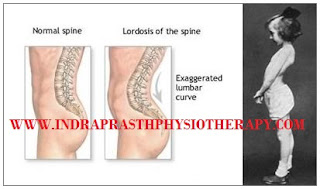
LORDOSIS - The term lordosis refers to the normal inward lordotic curvature of the lumbar and cervical regions of the spine. Excessive curvature of the lower back is known as lumbar hyperlordosis, commonly called hollow back. A major feature of lumbar hyperlordosis is a forward pelvic tilt, resulting in the pelvis resting on top of the thighs. Curvature in the opposite convex direction, in the thoracic and sacral regions is termed kyphotic. Lordosis is an increased inward curving of the lumbar spine. The spine has three types of curves: lordotic, kyphotic and scoliotic.
A small degree of both kyphotic and lordotic curvature is normal. Too much lordotic curving is called swayback (lordosis). Too much kyphotic curving causes round shoulders or hunched shoulders
Lordosis tends to make the buttocks appear more prominent. Children with hyperlordosis will have a large space underneath the lower back when lying face up on a hard surface. Some children have marked lordosis, but, most often fixes itself as the child grows. This is called benign juvenile lordosis. Spondylolisthesis may cause lordosis. In this condition, a bone in the spine slips out of the proper position onto the bone below it. You may be born with this. It can develop after certain sports activities, such as gymnastics. It may develop along with arthritis in the spine.


Lordosis symptoms

Symptoms of this abnormality depend upon the severity of the disease. Lordosis symptoms may include:
  • C-shape back when seen from a lateral aspect, with the buttocks being more prominent
  • A large gap between the lower back and the floor when lying on one’s back 
  • Pain and discomfort in the lower back.
  • Problems in moving in certain ways.