Saturday, 24 December 2016

CERVICAL SPONDYLOSIS

CERVICAL SPONDYLOSIS - Cervical spondylosis, also known as cervical osteoarthritis or neck arthritis, is a common, age-related condition that affects the joints and discs in your neck. It develops from wear and tear of the cartilage and bones found in your cervical spine, which is in your neck. While it’s largely due to age, it can be caused by other factors as well. The spinal disks can develop cracks, which allows leakage of the internal cushioning material. This material can press on the spinal cord and nerves, resulting in symptoms such as arm numbness and sciatica.
Factors other than aging can increase your risk of cervical spondylosis. These include:
- neck injuries
- work-related activities that put extra strain on your neck from heavy lifting
- holding your neck in an uncomfortable position for prolonged periods of time or repeating the same neck movements throughout the day (repetitive stress)
- genetic factors
- smoking
- being overweight and inactive


Most people with cervical spondylosis don’t have significant symptoms. If symptoms do occur, they can range from mild to severe and may develop gradually or occur suddenly One common symptom is pain around the shoulder blade. Patients will complain of pain along the arm and in the fingers. The pain might increase when:
  • standing
  • sitting
  • sneezing
  • coughing
  • tilting your neck backward


Thursday, 22 December 2016

PINCHED (COMPRESSED) NERVE

PINCHED (COMPRESSED) NERVE - Nerves extend from your brain and spinal cord, sending important messages throughout your body. If you have a pinched nerve your body may send you warning signals such as pain. Don't ignore these warning signals. Damage from a pinched nerve may be minor or severe. It may cause temporary or long-lasting problems. The earlier you get a diagnosis and treatment for nerve compression, the more quickly you'll find relief. In some cases, you can't reverse the damage from a pinched nerve. But treatment usually relieves pain and other symptoms.

A pinched nerve occurs when there is "compression" (pressure) on a nerve. The pressure may be the result of repetitive motions. Or it may happen from holding your body in one position for long periods, such as keeping elbows bent while sleeping. Nerves are most vulnerable at places in your body where they travel through narrow spaces but have little soft tissue to protect them. Nerve compression often occurs when the nerve is pressed between tissues such as:
-Ligament
-Tendon
-Bone


These symptoms may result from changes that develop in the spine's discs and bones. For example, if a disc weakens or tears -- known as a herniated disc -- pressure can get put on a spinal nerve.
Nerve compression in your neck or arm may also cause symptoms in areas such as your:
  • Elbow
  • Hand
  • Wrist
  • Fingers

Thursday, 17 November 2016

MALLET (BASEBALL) FINGER

MALLET FINGER - Mallet finger is an injury to the thin tendon that straightens the end joint of a finger or thumb. Although it is also known as "baseball finger," this injury can happen to anyone when an unyielding object strikes the tip of a finger or thumb and forces it to bend further than it is intended to go. As a result, you are not able to straighten the tip of your finger or thumb on your own.
Tendons are tissues that connect muscles to bone. The muscles that move the fingers and thumb are located in the forearm. Long tendons extend from these muscles through the wrist and attach to the small bones of the fingers and thumb. The extensor tendons on the top of the hand straighten the fingers. The flexor tendons on the palm side of the hand bend the fingers.

In a mallet injury, when an object hits the tip of the finger or thumb, the force of the blow tears the extensor tendon. Occasionally, a minor force such as tucking in a bed sheet will cause a mallet finger.
The injury may rupture the tendon or pull the tendon away from the place where it attaches to the finger bone (distal phalanx). In some cases, a small piece of bone is pulled away along with the tendon. This is called an avulsion injury. The finger is usually painful, swollen, and bruised. The fingertip will droop noticeably and will straighten only if you push it up with your other hand.


Mallet finger can be caused by a blunt force on the DIP. Patients who are diagnosed with mallet finger have an inability to extend their finger and experience pain and numbness. Depending on how severe the injury is, the patient can be prescribed medication in order to prevent infection. Also, most mallet finger injuries can be treated without surgery.

Tuesday, 15 November 2016

LOW BACK PAIN

LOW BACK PAIN - Low back pain is a universal human experience -- almost everyone has it at some point. The lower back, which starts below the ribcage, is called the lumbar region. Pain here can be intense and is one of the top causes of missed work. Fortunately, low back pain often gets better on its own. When it doesn't, there are effective treatments. In most episodes of low back pain, a specific underlying cause is not identified or even looked for, with the pain believed to be due to mechanical problems such as joint strain. If the pain does not go away with conservative treatment or if it is accompanied by "red flags" such as unexplained weight loss, fever, or significant problems with feeling or movement, further testing may be needed to look for a serious problem.

Lower back pain can be caused by a variety of problems with any parts of the complex, interconnected network of spinal muscles, nerves, bones, discs or tendons in the lumbar spine. Typical sources of low back pain include:
  • The large nerve roots in the low back that go to the legs may be irritated
  • The smaller nerves that supply the low back may be irritated
  • The large paired lower back muscles may be strained
  • The bones, ligaments or joints may be damaged
  • An intervertebral disc may be degenerating


Certain causes of lower back pain have a tendency to occur more often in younger individuals versus older adults:
  • Younger adults (30 to 60 year olds) are more likely to experience back pain from the disc space itself or from a back muscle strain or other soft tissue strain.
  • Older adults (over 60) are more likely to suffer from pain related to joint degeneration or from a compression fracture.    

Saturday, 12 November 2016

TOTAL HIP REPLACEMENT

TOTAL HIP REPLACEMENT - A total hip replacement is a surgical procedure whereby the diseased cartilage and bone of the hip joint is surgically replaced with artificial materials. The normal hip joint is a ball and socket joint. The socket is a "cup-shaped" component of the pelvis called the acetabulum. The ball is the head of the thighbone. Total hip joint replacement involves surgical removal of the diseased ball and socket and replacing them with a metal ball and stem inserted into the femur bone and an artificial plastic cup socket. The metallic artificial ball and stem are referred to as the "femoral prosthesis" and the plastic cup socket is the "acetabular prosthesis." Upon inserting the prosthesis into the central core of the femur, it is fixed with a bony cement called methylmethacrylate. Alternatively, a "cementless" prosthesis is used that has microscopic pores which allow bony ingrowth from the normal femur into the prosthesis stem. This "cementless" hip is felt to have a longer duration and is considered especially for younger patients. Total hip replacement is also referred to as total hip arthroplasty.


Total hip replacements are performed most commonly because of progressively worsening of severe arthritis in the hip joint. The most common type of arthritis leading to total hip replacement is degenerative arthritis of the hip joint. This type of arthritis is generally seen with aging, congenital abnormality of the hip joint. The progressively intense chronic pain, together with impairment of daily function including walking climbing stairs, and even arising from a sitting position, eventually become reasons to consider a total hip replacement. Because replaced hip joints can fail with time, whether and when to perform total hip replacement are not easy decisions, especially in younger patients.

Monday, 7 November 2016

TOTAL KNEE REPLACEMENT

TOTAL KNEE REPLACEMENT - Patients with severe destruction of the knee joint associated with progressive pain and impaired function maybe candidates for total knee replacement. Osteoarthritis is the most common reason for knee replacement operation. Physical therapy is an essential part of rehabilitation after total knee replacement. A TKR is a complex procedure that requires an orthopedic surgeon to make precise measurements and skillfully remove the diseased portions of your bone, in order to shape the remaining bone to accommodate the knee implant. After knee replacement surgery, you are usually sent home or to a rehabilitation facility, depending on your condition at that time. If you are sent to a facility, the average rehabilitation stay is approximately seven to ten days.

After knee replacement surgery, you should not pivot or twist on the involved leg for at least six weeks. Also during this time, when lying in bed, you should keep the involved knee as straight as possible. Kneeling and squatting also should be avoided soon after knee joint replacement surgery. Stair climbing should be kept to a minimum. Make the necessary arrangements so that you will only have to go up and down the steps once or twice a day. A firm, straight-back chair is extremely helpful in adhering to these joint precautions. Recliners should not be used. To help avoid falls, all throw rugs should be removed from the floor and rooms should be kept free of unnecessary debris. Enthusiastic pets should be kept far away until you have healed.


Tuesday, 25 October 2016

LOWER CROSSED SYNDROME

Lower-Crossed Syndrome (LCS) - . In LCS, tightness of the thoracolumbar extensors on the dorsal side crosses with tightness of the iliopsoas and rectus femoris. Weakness of the deep abdominal muscles ventrally crosses with weakness of the gluteus maximus and medius. This pattern of imbalance creates joint dysfunction, particularly at the L4-L5 and L5-S1 segments, SI joint, and hip joint. Specific postural changes seen in LCS include anterior pelvic tilt, increased lumbar lordosis, lateral lumbar shift, lateral leg rotation, and knee hyperextension. If the lordosis is deep and short, then imbalance is predominantly in the pelvic muscles; if the lordosis is shallow and extends into the thoracic area, then imbalance predominates in the trunk muscles.


Causes

  • Prolonged sitting, particularly with bad posture
  • Physical inactivity
  • Regular performance of sports and activities that involve an uneven stimulation of the muscles that are involved in LCS
  • Poor exercise technique.
  • Imbalanced strength training
  • Genetic predispositions
 The development of LCS initiates a vicious cycle. Because the gluteals and abdominals are weak, their function is compromised, and other muscles such as the hamstrings and lower back muscles are recruited to assist them in performing activities such as walking, running, and squatting. This leads to overuse and tightness of the hamstrings and lower back muscles, and a further weakening of the abdominals and gluteals.

Wednesday, 5 October 2016

LORDOSIS

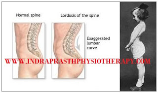
LORDOSIS - The term lordosis refers to the normal inward lordotic curvature of the lumbar and cervical regions of the spine. Excessive curvature of the lower back is known as lumbar hyperlordosis, commonly called hollow back. A major feature of lumbar hyperlordosis is a forward pelvic tilt, resulting in the pelvis resting on top of the thighs. Curvature in the opposite convex direction, in the thoracic and sacral regions is termed kyphotic. Lordosis is an increased inward curving of the lumbar spine. The spine has three types of curves: lordotic, kyphotic and scoliotic.
A small degree of both kyphotic and lordotic curvature is normal. Too much lordotic curving is called swayback (lordosis). Too much kyphotic curving causes round shoulders or hunched shoulders
Lordosis tends to make the buttocks appear more prominent. Children with hyperlordosis will have a large space underneath the lower back when lying face up on a hard surface. Some children have marked lordosis, but, most often fixes itself as the child grows. This is called benign juvenile lordosis. Spondylolisthesis may cause lordosis. In this condition, a bone in the spine slips out of the proper position onto the bone below it. You may be born with this. It can develop after certain sports activities, such as gymnastics. It may develop along with arthritis in the spine.


Lordosis symptoms

Symptoms of this abnormality depend upon the severity of the disease. Lordosis symptoms may include:
  • C-shape back when seen from a lateral aspect, with the buttocks being more prominent
  • A large gap between the lower back and the floor when lying on one’s back 
  • Pain and discomfort in the lower back.
  • Problems in moving in certain ways.

Tuesday, 27 September 2016

KYPHOSIS

KYPHOSIS - Kyphosis refers to the abnormally excessive convex kyphotic curvature of the spine as it occurs in the cervical, thoracic and sacral regions. Kyphosis can be called roundback. It can result from degenerative diseases such as arthritis, developmental problems, most commonly, osteoporosis with compression fractures of the vertebra, Multiple myeloma or trauma. A normal thoracic spine extends from the 1st to the 12th vertebra and should have a slight kyphotic angle, ranging from 20° to 45°. When the "roundness" of the upper spine increases past 45° it is called kyphosis or "hyperkyphosis".


Kyphosis is a forward rounding of the back. Some rounding is normal, but the term "kyphosis" usually refers to an exaggerated rounding of the back. While kyphosis can occur at any age, it's most common in older women. Age-related kyphosis often occurs after osteoporosis weakens spinal bones to the point that they crack and compress. Other types of kyphosis are seen in infants or teens due to malformation of the spine or wedging of the spinal bones over time. Mild kyphosis causes few problems, but severe cases can cause pain and be disfiguring. Treatment for kyphosis depends on your age, the cause of the curvature and its effects.The spine has a series of normal curves when viewed from the side. These curves help to better absorb the loads applied to the spine from the weight of the body. The cervical spine and lumbar spine are have a normal inward curvature that is medically referred to as lordosisor "lordotic" curvature by which the spine is bent backward. The thoracic spine has a normal outward curvature that is medically referred to as kyphosis or the "kyphotic" curve by which the spine is bent forward. In this discussion, the term kyphosis will be used to discuss abnormal kyphosis.

Wednesday, 21 September 2016

JUMPERS KNEE

JUMPERS KNEE - Jumpers knee or patellar tendonitis is an overuse injury that results in pain at the front of the knee, localised at a point towards the bottom of the kneecap. Repetitive strain from too much running or jumping causes inflammation or degeneration of the patella tendon. Patellar tendonitis can be a tricky condition to treat and requires a substantial period of rest and a thorough treatment and rehabilitation program. In the most severe or persistent cases, surgery may be required. Jumper's knee also known as patellar tendonitis or patellar tendinopathy. It is an inflammation or injury of the patellar tendon, the cord-like tissue that joins the patella to the tibia. Jumper's knee is an overuse injury. Constant jumping, landing, and changing direction can cause strains, tears, and damage to the patellar tendon. Who regularly play sports that involve a lot of repetitive jumping — like track and field, basketball, volleyball, gymnastics, running, and soccer can put a lot of strain on their knees. Jumper's knee can seem like a minor injury that isn't really that serious. Because of this, many athletes keep training and competing and tend to ignore the injury or attempt to treat it themselves. But it's important to know that jumper's knee is a serious condition that can get worse over time and ultimately require surgery. Early medical attention and treatment can help prevent continued damage to the knee.


Symptoms of jumpers knee consist of pain at the bottom front of the kneecap over what is called the lower pole of the patella. The bottom of the patella will be very tender when pressing in and may appear larger or thicker than the unaffected side. The athlete is likely to experience aching and stiffness after exercise and pain when contracting the quadriceps muscles in acute cases. In particular jumping activities are likely to cause most pain or discomfort.

The patella tendon, also known as the patella ligament joins the kneecap or patella to the shin bone or tibia. The large quadriceps muscles at the front of the thigh pull on the kneecap producing huge forces through the patella tendon, especially when jumping. Jumpers knee is an overuse injury. With repeated strain, micro-tears as well as collagen degeneration may occur in the tendon. This is known as patellar tendinopathy or Jumpers Knee

Saturday, 17 September 2016

JOINT PAIN

JOINT PAIN - Joint pain can range from mildly irritating to debilitating. It may go away after a few weeks (acute), or last for several weeks or months (chronic). Even short-term pain and swelling in the joints can affect your quality of life. Joint Pain is extremely common. In one national survey, about one-third of adults reported having joint pain within the past 30 days. Knee Pain was the most common complaint, followed by shoulder and hip pain, but joint pain can affect any part of your body, from your ankles and feet to your shoulders and hands. As you get older, painful joints become increasingly more common. Joint pain can be caused by injury affecting any of the ligaments, bursae, or tendons surrounding the joint. Injury can also affect the ligaments, cartilage, and bones within the joint.

Symptoms and signs associated with joint pain can include:
  • joint swelling,
  • joint tenderness,
  • joint warmth,
  • locking of the joint,
  • loss of range of motion of the joint,
  • stiffness,
  • joint redness
  • weakness
  • limping


 Joint pain can be aggravated by motion, pressure, or weight-bearing resistance with activity. Joint pain can be associated with local warmth, swelling, and tenderness. Joint pain can be caused by injury or disease of the joint or adjacent tissues. A joint is the area at which two bone ends meet to provide motion to a body part. A typical joint is composed of bones that are separated by cartilage that serves as cushioning pad for the adjacent bones. Ligaments attach bone to bone around the joint. Bursae are fluid-filled sacs that provide a gliding surface for adjacent tendons. Tendons attach muscle to bone around the joint. Injury or disease to any of the structures of the joint can lead to pain in the joint. Joint pain is also referred to as arthralgia.

Friday, 16 September 2016

JERSEY FINGER

JERSEY FINGER - Jersey Finger represents rupture of the Flexor Digitorum Profundus. This usually happens when an athlete grabbing an opponent's shirt undergoes forced extension of the DIP while it is flexed. For this reason, it is called "grasping jersey finger" or "rugby finger". The ring finger is involved more often. The rupture of the FDP tendon from its insertion on the distal phalanx known as Jersey Finger is often misdiagnosed as a sprained or jammed finger, as there is no characteristic deformity associated with it.

The injury is typically caused by forceful passive extension while the flexor digitorum profundus muscle is contracting. A common example is in football when the flexed finger is caught in a jersey while the athlete is attempting to make a tackle; hence the term jersey finger.Jersey finger symptoms include pain in the finger tip and inability to bend the finger normally although it can still be forced into a bended position. Tenderness on the pad of the finger will be present along with swelling and bruising which may develop later in the finger tip. It may be possible to feel the tendon as a bunched up soft mass on the palm side of the hand.


  • A pop or rip felt in the finger at the time of the injury
  • Tenderness, swelling and warmth of the injured finger
  • Pain when moving the injured finger and the inability to bend the last joint
  • Occasionally a lump felt in the palm of the finger
  • Bruising after 48 hours

Tuesday, 13 September 2016

IT BAND SYNDROME

IT BAND SYNDROME - Iliotibial band syndrome is one of the leading causes of lateral knee pain in runners. The iliotibial band is a thick band of fascia on the lateral aspect of the knee, extending from the outside of the pelvis, over the hip and knee, and inserting just below the knee. The band is crucial to stabilizing the knee during running, as it moves from behind the femur to the front of the femur during activity. The continual rubbing of the band over the lateral femoral epicondyle, combined with the repeated flexion and extension of the knee during running may cause the area to become inflamed. The iliotibial band is a thick band that begins at the iliac crest in the pelvis, runs down the lateral part of the thigh, and crosses the knee to attach into the top part of the tibia or shinbone. The iliotibial (IT) band helps stabilize the outside part of the knee through its range of motion. Iliotibial band syndrome is the most common cause of lateral knee pain in runners and bicyclists.

Prevention that includes maintaining flexibility and strength of the low back, hips, knees, and leg muscles is key to avoiding recurrence. Inflammation of the iliotibial band can occur as it travels back and forth, crossing the bony prominence of the femoral epicondyle as the knee flexes and extends. The iliotibial band runs along the lateral or outside aspect of the thigh, from the pelvis to the tibia, crossing both the hip and knee joints. The iliotibial band is an important stabilizer structure of the lateral part of the knee as the joint flexes and extends.


 Pain may be felt when bending and straightening the knee which may be made worse by pressing in at the side of the knee over the sore part. There might be tightness in the iliotibial band which runs down the outside of the thigh. Weakness in hip abduction or moving the leg out sideways is another common sign. Tender trigger points in the gluteal muscles or buttocks area may also be present.

Friday, 2 September 2016

HERNIATED DISK

HERNIATED DISK - A herniated disk refers to a problem with one of the rubbery cushions (disks) between the individual bones (vertebrae) that stack up to make your spine. A spinal disk is a little like a jelly donut, with a softer center encased within a tougher exterior. Sometimes called a slipped disk or a ruptured disk, a herniated disk occurs when some of the softer "jelly" pushes out through a crack in the tougher exterior. A herniated disk can irritate nearby nerves and result in pain, numbness or weakness in an arm or leg. On the other hand, many people experience no symptoms from a herniated disk. There are many terms used to describe spinal disc pathology and associated pain, such as "herniated disc," "pinched nerve," and "bulging disc," and all are used differently and, at times, interchangeably.


Herniated disc facts

  • The discs are pads that serve as "cushions" between the vertebral bodies, which minimize the impact of movement on the spinal column.
  • Each disc is designed like a jelly donut with a central softer component (nucleus pulposus).
  • Abnormal rupture of the central portion of the disc is referred to as a disc herniation.
  • The most common location for a herniated disc to occur is in the disc at the level between the fourth and fifth lumbar vertebrae in the low back.

Sometimes called a slipped or ruptured disk, a herniated disk most often occurs in your lower back. It is one of the most common causes of low back pain, as well as leg pain (sciatica). Between 60% and 80% of people will experience low back pain at some point in their lives. A high percentage of people will have low back and leg pain caused by a herniated disk. Although a herniated disk can sometimes be very painful, most people feel much better with just a few weeks or months.

Wednesday, 31 August 2016

HEMIPLEGIA

HEMIPLEGIA - Hemiplegia is a condition that affects one side of the body. We talk about a right or left hemiplegia, depending on the side affected. It is caused by injury to parts of the brain that control movements of the limbs, trunk, face, etc. This may happen before, during or soon after birth, when it is known as congenital hemiplegia, or later in life as a result of injury or illness, in which case it is called acquired hemiplegia. Generally, injury to the left side of the brain will cause a right hemiplegia and injury to the right side a left hemiplegia. The causes of congenital hemiplegia are mostly unknown, and usually parents only become aware of their child’s hemiplegia during infancy or early childhood as the child’s difficulty with movement on one side gradually becomes obvious. There is a higher risk in very premature babies and with multiple pregnancies, and it is unclear whether a difficult birth may be an occasional factor.


Injury to the brain cells that control movements in one half of the body cause hemiplegia. Hence, symptoms largely depend upon the part of the brain affected. The same can be said about the severity of individual symptoms.
  • Difficulty in walking.
  • Problems in balance, losses balance when trying to walk
  • Difficulty in swallowing
  • Trouble with vision. Blurred vision or weakness of the eyes.
  • Speech becomes difficult.
  • Numbness, tingling or loss of sensations on one half of the body.
  • Loss of control over bladder and bowel movements leading to an inability to hold on to stool or urine.
  • Unable to perform tasks like holding objects, tying laces, dressing oneself, buttoning etc.
  • Feeling depressed
  • Heightened emotional sensitivity with inability to handle stressful situations.
  • Memory seems poor. Unable to recall recent or past events concerning people, places and activities.

'Hemiplegia' means complete paralysis of one half of the body, including one arm and leg. Any disease or injury in the motor centers of the brain can cause hemiplegia. Hemiplegia is a more severe form of 'hemiparesis' wherein one half of the body is only weakened. It is also very different from the conditions of paraplegia & quadriplegia, which are commonly confused with hemiplegia. Stroke: is the commonest cause of hemiplegia. Insufficient blood supply to the brain leads to loss of brain functions. A clot formed within the blood vessel blocking the blood supply'. A thrombus breaks away from its site of origin and forms a block elsewhere in the circulation. A bleed from a blood vessel supplying the brain.

Saturday, 27 August 2016

HEEL PAIN

HEEL PAIN - Heel pain is a common foot condition. It's usually felt as an intense pain when using the affected heel. Heel pain usually builds up gradually and gets worse over time. The pain is often severe and occurs when you place weight on your heel. The pain is usually worse first thing in the morning, or when you first take a step after a period of inactivity. Walking usually improves the pain, but it often gets worse again after walking or standing for a long time. Some people may limp or develop an abnormal walking style as they try to avoid placing weight on the affected heel.
The pain is usually worse first thing in the morning, or when you first take a step after a period of inactivity. Walking usually improves the pain, but it often gets worse again after walking or standing for a long time. Some people may limp or develop an abnormal walking style as they try to avoid placing weight on the affected heel.


Heel pain has a number of causes that are typically associated with overuse of the heel bone. You can strain your heel by pounding your feet on hard surfaces, being overweight, or wearing shoes that do not fit properly. These strains can irritate the heel’s bones, muscles, or tendons. Other common causes of heel pain include the following conditions. our foot and ankle are made up of 26 bones, 33 joints, and more than 100 tendons. The heel is the largest bone in your foot. If you overuse or injure your heel, you may experience heel pain. This can range from mild to disabling. In many cases, if you have heel pain, you will need a doctor or podiatrist to diagnose the cause.


There are two different categories of heel pain. The first is caused by over-use repetitive stress which refers to a soreness resulting from too much impact on a specific area of the foot. This condition, often referred to as "heel pain syndrome," can be caused by shoes with heels that are too low, a thinned out fat pad in the heel area, or from a sudden increase in activity. Plantar fasciitis, a very common diagnosis of heel pain, is usually caused from a biomechancial problem, such as over-pronation (flat feet). The plantar fascia is a broad band of fibrous tissue that runs along the bottom surface of the foot, from the heel through the midfoot and into the forefoot. Over-pronation can cause the plantar fascia to be excessively stretched and inflamed, resulting in pain in the heel and arch areas of the foot. Often the pain will be most intense first thing in the morning or after a prolonged period of rest. The pain will gradually subside as the day progresses.

Monday, 22 August 2016

HALLUX VALGUS

HALLUX VALGUS - A hallux valgus deformity, commonly called a bunion, is when there is medial deviation of the first metatarsal and lateral deviation of the great toe (hallux). The condition can lead to painful motion of the joint and shoe wear difficulty. It is important for a treating physician to understand the pathogenesis and surgical treatment options to correct hallux valgus deformities to provide the utmost care for patients with this painful forefoot deformity. Hallux valgus is a common, painful orthopaedic foot and ankle deformity. Most hallux valgus deformities can be treated conservatively with appropriate shoewear modifications, orthotics, and bunion splints. Surgery is indicated for pain relief and appropriate counseling of patients and their expectations are essential for a successful outcome. It is important to understand the pathogenesis of a hallux valgus deformity because surgical treatment options are based on the clinical examination findings. Appropriate surgical management results in improvement in the patient’s pain and overall function.​ The toe tilts over towards the smaller toes and a bony lump appears on the inside of the foot. Sometimes a soft fluid swelling develops over the bony lump. The bony lump is the end of the "knuckle-bone" of the big toe which becomes exposed as the toe tilts out of place.


 Many people do not experience symptoms in the early stages of bunion formation. Symptoms are often most noticeable when the bunion gets worse and with certain types of footwear. These include shoes that crowd the toes and/or high-heeled shoes. When symptoms do occur, they may include:

    • Physical discomfort or pain
    • A burning feeling
    • Redness and swelling
    • Possible numbness
    • Difficulty walking
     

Saturday, 20 August 2016

GOUT

GOUT - Gout is a kind of arthritis. It can cause an attack of sudden burning pain, stiffness, and swelling in a joint, usually a big toe. These attacks can happen over and over unless gout is treated. Over time, they can harm your joints, tendons, and other tissues. Gout is most common in men.Gout is caused by too much uric acid in the blood. Most of the time, having too much uric acid isn't harmful. Many people with high levels in their blood never get gout. But when uric acid levels in your blood are too high, the uric acid may form hard crystals in your joints.


The most common sign of gout is a nighttime attack of swelling, tenderness, redness, and sharp pain in your big toe. You can also get gout attacks in your foot, ankle, or knees, or other joints. The attacks can last a few days or many weeks before the pain goes away. Gout is characterized by sudden, severe attacks of pain, redness and tenderness in joints, often the joint at the base of the big toe. Gout — a complex form of arthritis — can affect anyone. Men are more likely to get gout, but women become increasingly susceptible to gout after menopause. An attack of gout can occur suddenly, often waking you up in the middle of the night with the sensation that your big toe is on fire. The affected joint is hot, swollen and so tender that even the weight of the sheet on it may seem intolerable.


Gout, a painful form of arthritis, occurs when high levels of uric acid in the blood cause crystals to form and accumulate around a joint. Uric acid is produced when the body breaks down a chemical called purine. Purine occurs naturally in your body, but it's also found in certain foods. Uric acid is eliminated from the body in urine. A gout diet may help decrease uric acid levels in the blood. While a gout diet is not a cure, it may lower the risk of recurring painful gout attacks and slow the progression of joint damage. Medication also is needed to manage pain and to lower levels of uric acid.

Wednesday, 17 August 2016

GOLFER'S ELBOW

GOLFER'S ELBOW - Golfer's elbow is a condition that causes pain where the tendons of your forearm muscles attach to the bony bump on the inside of your elbow. The pain might spread into your forearm and wrist.
Golfer's elbow is similar to tennis elbow, which occurs on the outside of the elbow. It's not limited to golfers. Tennis players and others who repeatedly use their wrists or clench their fingers also can develop golfer's elbow.

The pain of golfer's elbow can come on suddenly or gradually. The pain might worsen when you:
  • Swing a golf club or racket
  • Squeeze or pitch a ball
  • Shake hands
  • Turn a doorknob
  • Lift weights
  • Pick up something with your palm down
  • Flex your wrist
The condition is called Golfer's Elbow because in making a golf swing this tendon is stressed, especially if a non-overlapping grip is used; many people, however, who develop the condition have never handled a golf club. It is also sometimes called Pitcher's Elbow due to the same tendon being stressed by the throwing of objects such as a baseball, but this usage is much less frequent.


Golfer's elbow is also known as medial epicondylitis and causes pain and inflammation at the point where the flexor tendons of the forearm are attached to the upper arm. The pain is focused on the bony bump on the inside of the elbow and may radiate along the forearm. Golfer's elbow is usually caused by overuse of the muscles in the forearm that allow you to rotate your arm and flex your wrist. Repetitive flexing, gripping or swinging can cause pulls or tiny tears in the tendons close to where they are attached to the bone.

Tuesday, 16 August 2016

FROZEN SHOULDER


FROZEN SHOULDER - In frozen shoulder, the shoulder capsule thickens and becomes tight. Stiff bands of tissue — called adhesions — develop. In many cases, there is less synovial fluid in the joint.
The hallmark sign of this condition is being unable to move your shoulder - either on your own or with the help of someone else. Pain from frozen shoulder is usually dull or aching. It is typically worse early in the course of the disease and when you move your arm. The pain is usually located over the outer shoulder area and sometimes the upper arm. Frozen shoulder (adhesive capsulitis) is stiffness, pain, and limited range of movement in your shoulder. It may happen after an injury or overuse or from a disease such as stroke. The tissues around the joint stiffen, scar tissue forms, and shoulder movements become difficult and painful. The condition usually comes on slowly, then goes away slowly over the course of a year or more.

 The bones, ligaments and tendons that make up your shoulder joint are encased in a capsule of connective tissue. Frozen shoulder occurs when this capsule thickens and tightens around the shoulder joint, restricting its movement. The shoulder has a spheroidal joint, in which the round part of one bone fits into the concavity of another. The proximal humerus fits into socket of the scapula. Frozen shoulder is thought to cause the formation of scar tissue in the shoulder, which makes the shoulder joint's capsule thicken and tighten, leaving less room for movement. Therefore, movement may be stiff and even painful.


• CHARACTERISED BY PAINFUL & STIFF GH JOINT D/T LOSS OF RESILIENCE OF JOINT CAPSULE , POSSIBLY WITH ADHESIONS B/W ITS SYNOVIAL FOLDS.
• DENSE ADHESIONS, CAPSULAR THICKENING, CAPSULAR RESTRICTIONS IN FOLDS OF JOINT CAPSULE
• GLOBAL LOSS OF ACTIVE & PASSIVE GH MOTION.
• LOSS OF ER *HALLMARK*

Saturday, 6 August 2016

FIBROMYALGIA


FIBROMYALGIA - Fibromyalgia is a disorder characterized by widespread musculoskeletal pain accompanied by fatigue, sleep, memory and mood issues. Researchers believe that fibromyalgia amplifies painful sensations by affecting the way your brain processes pain signals. Women are much more likely to develop fibromyalgia than are men. Many people who have fibromyalgia also have tension headaches, temporomandibular joint (TMJ) disorders, irritable bowel syndrome, anxiety and depression. Other symptoms include feelind tired to a degree that normal activities are affected, sleep problems, and troubles with memory. Some people also report restless legs syndrome, bowel or bladder problems, numbness and tingling, and sensitivity to noise, lights or temperature. Fibromyalgia is frequently associated with depression and anxiety. Other types of chronic pain are also frequently present.

The treatment of fibromyalgia can be difficult. Recommendations often include getting enough sleep, exercising regularly, and eating ahealthy diet. Cognitive behavioral therapy may also be helpful. Use of opioid pain medication is controversial with some stating they are poorly supported and others saying that weak opioids may be reasonable if other medications are not effective.


Symptoms of fibromyalgia include:
  • Chronic muscle pain, muscle spasms, or tightness
  • Moderate or severe fatigue and decreased energy
  • Insomnia or waking up feeling just as tired as when you went to sleep
  • Stiffness upon waking or after staying in one position for too long
  • Difficulty remembering, concentrating, and performing simple mental task.
  • Abdominal pain, bloating, nausea, and constipation alternating with diarrhea
  • Tension or migraine headaches
  • Jaw and facial tenderness
  • Sensitivity to one or more of the following: odors, noise, bright lights, medications, certain foods, and cold
  • Feeling anxious or depressed
  • Numbness or tingling in the face, arms, hands, legs, or feet
  • Increase in urinary urgency or frequency.
  • Reduced tolerance for exercise and muscle pain after exercise
  • A feeling of swelling in the hands and feet

Friday, 5 August 2016

ERB'S PALSY

ERB'S PALSY - Erb's palsy is a paralysis of the arm caused by injury to the upper group of the arm's main nerves, specifically the severing of the upper trunk C5-C6 nerves. These form part of the brachial plexus, comprising the ventral rami of spinal nerves C5–C8 and thoracic nerve. The paralysis can be partial or complete; the damage to each nerve can range from bruising to tearing. The most commonly involved root is C5 as this is mechanically the furthest point from the force of traction, therefore, the first/most affected. Erb–Duchenne palsy presents as a lower motor neuron syndrome associated with sensibility disturbance and vegetative phenomena.

The signs of Erb's Palsy include loss of sensation in the arm and paralysis and atrophy of the deltoid, biceps, and brachialis muscles. "The position of the limb, under such conditions, is characteristic: the arm hangs by the side and is rotated medially; the forearm is extended and pronated. The arm cannot be raised from the side; all power of flexion of the elbow is lost, as is also supination of the forearm". The resulting biceps damage is the main cause of this classic physical position commonly called "waiter's tip."


The brachial plexus is a network of nerves near the neck that give rise to all the nerves of the arm. These nerves provide movement and feeling to the shoulder, arm, hand, and fingers. Palsy means weakness, and brachial plexus birth palsy causes arm weakness and loss of motion.

 Symptoms
  • Weakness in one arm
  • Loss of feeling in the arm
  • Partial or total paralysis of the arm

Thursday, 4 August 2016

DIABETIC NEUROPATHY

DIABETIC NEUROPATHY - Diabetic neuropathy is a type of nerve damage that can occur if you have diabetes. High blood sugar can injure nerve fibers throughout your body, but diabetic neuropathy most often damages nerves in your legs and feet.Depending on the affected nerves, symptoms of diabetic neuropathy can range from pain and numbness in your extremities to problems with your digestive system, urinary tract, blood vessels and heart.Peripheral neuropathy is the most common form of diabetic neuropathy. Your feet and legs are often affected first, followed by your hands and arms. Neuropathy is damage to nerves, and diabetic neuropathy is damage to nerves that occurs as a result of diabetes. Diabetes is thought to damage nerves as a result of prolonged elevated levels of blood glucose. Diabetic neuropathy can affect different parts of the body, and symptoms can range from mild to severe. Diabetic neuropathy is the most common complication of diabetes.

SIGN AND SYMPTOMS
  • Numbness or reduced ability to feel pain or temperature changes
  • A tingling or burning sensation
  • Sharp pains or cramps
  • Increased sensitivity to touch
  • Muscle weakness
  • Loss of reflexes in the ankle
  • Loss of balance and coordination
  • Serious foot problems, such as ulcers, infections, deformities, and bone and joint pain.


 About 60 to 70 percent of people with diabetes have some form of neuropathy. People with diabetes can develop nerve problems at any time, but risk rises with age and longer duration of diabetes. The highest rates of neuropathy are among people who have had diabetes for at least 25 years. Diabetic neuropathies also appear to be more common in people who have problems controlling their blood glucose, also called blood sugar, as well as those with high levels of blood fat and blood pressure and those who are overweight.

Wednesday, 3 August 2016

DE QUERVAIN'S TENDINOSIS


DE QUERVAIN'S TENDINOSIS - De Quervain's tendinosis occurs when the tendons around the base of the thumb are irritated or constricted. The word "tendinosis" refers to a swelling of the tendons. Swelling of the tendons, and the tendon sheath, can cause pain and tenderness along the thumb side of the wrist. This is particularly noticeable when forming a fist, grasping or gripping something, or when turning the wrist.

Symptoms of de Quervain's tenosynovitis include:
  • Pain near the base of your thumb
  • Swelling near the base of your thumb
  • Difficulty moving your thumb and wrist when you're doing something that involves grasping or pinching
  • A "sticking" or "stop-and-go" sensation in your thumb when moving it
Patients often describe a feeling of inflammation, but studies have shown that the process is not inflammatory.  People of all ages get it.  When new mothers develop de Quervain syndrome, it typically appears 4 to 6 weeks after delivery.  The old theory that it was caused by wringing out cloth diapers has been replaced by concerns about holding the baby, but changes in hormones and swelling seem more probable.


Signs
Pain may be felt over the thumb side of the wrist. This is the main symptom. The pain may appear either gradually or suddenly. Pain is felt in the wrist and can travel up the forearm. The pain is usually worse when the hand and thumb are in use. This is especially true when forcefully grasping objects or twisting the wrist.
  • Swelling may be seen over the thumb side of the wrist. This swelling may accompany a fluid-filled cyst in this region.
  • A "catching" or "snapping" sensation may be felt when moving the thumb.
  • Pain and swelling may make it difficult to move the thumb and wrist.